Credit: This article was first published on the Motic Europe / Americas Web site
Basics of Light Microscopy: Incident Light Microscopy for opaque samples (I)
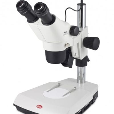
Motic Panthera BF/DF Met Microscope – MMS Microscopes
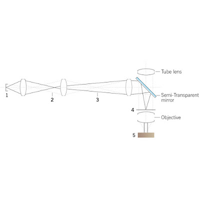
Light Path of Incident Light Microscope – MMS Microscopes
1Light source
Met Microscope Field Diaphragm
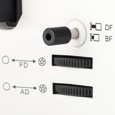
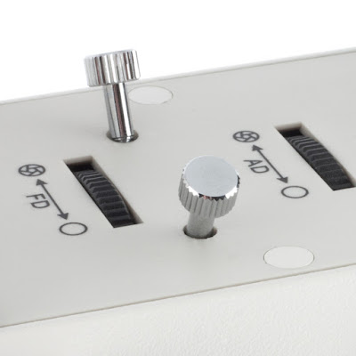
Brightfield BF & Darkfield DF Control of Incident Light Microscope – MMS Microscopes
The Field diaphragm defines the illuminated area, the aperture diaphragm regulates contrast as well as depth of field. In case of a flat polished sample surface, depth of field of course is negligible.
The semi-transparent mirror in the front part of any Epi-illuminator is a funny thing. It deflects the light by 45°, at the same time being transparent by 50%. In a Fluorescence setup, a dichroic mirror separates different colors by wave length quality, here we talk about a 50/50 quantitative separation of white light.
Epi-illumination in a compound microscope can be verified in 2 different hardware setups. The Inverted microscope is a more expensive and complex construction but has got obvious advantages. First, we need only one flat surface on the sample to start the investigation. Secondly, we do enjoy the freedom of an enlarged sample size. As in an Inverted metallurgical microscope mostly the focus mechanism is done by moving the nosepiece with its objectives, samples with reduced weight limitations can be handled.
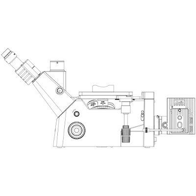
Inverted Metallurgical Met Lab Microscope from MMS Microscopes
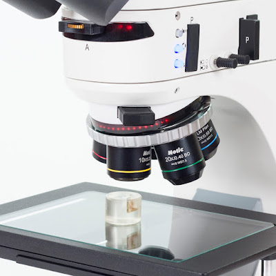
Met Materials Microscope with Brightfield (BF) Darkfield (DF) Met Objectives
For upright microscopes, we need 2 flat surfaces orientated parallel for the positioning of the sample.
Motic Incident Light Met Microscope from MMS Microscopes
A mechanical press together with some modeling clay from a toy shop are helpful tools to make a metallurgical sample ready for investigation. A metal slide is the carrier, and the flexible modeling clay with the sample placed on top can be pressed to position the sample properly.
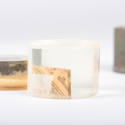
Met Lab sample prep
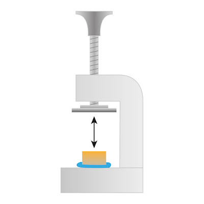
Met Lab Microscope Sample Press from MMS microscopes
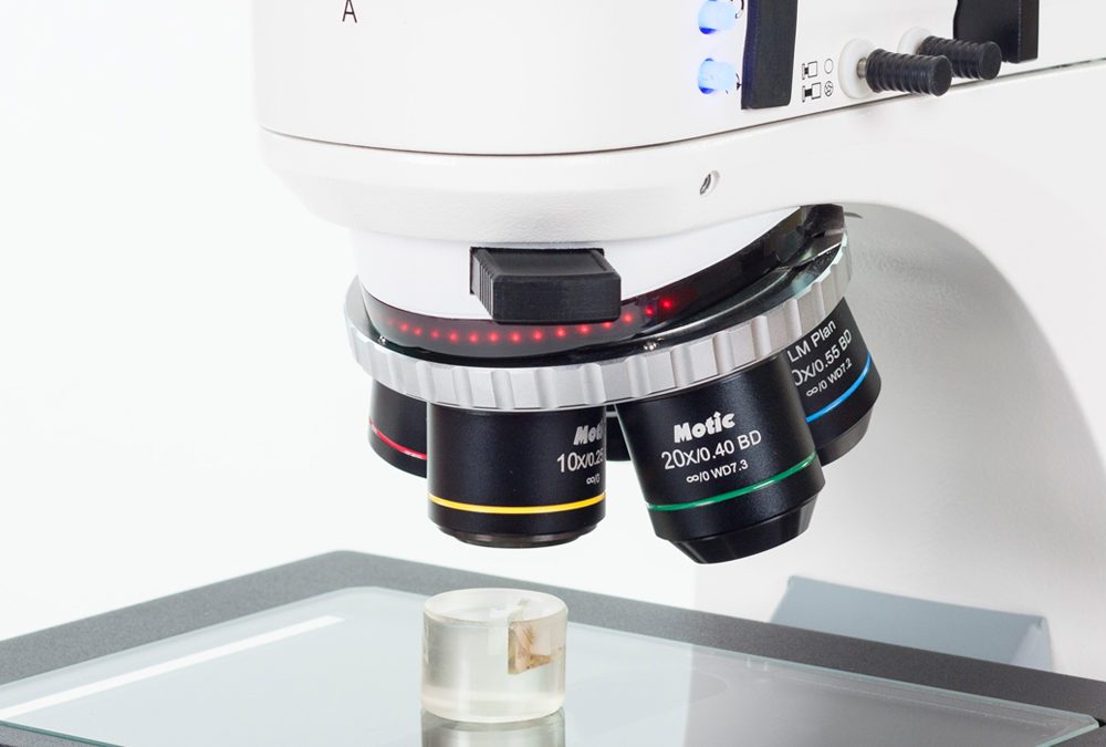
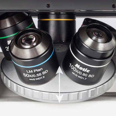
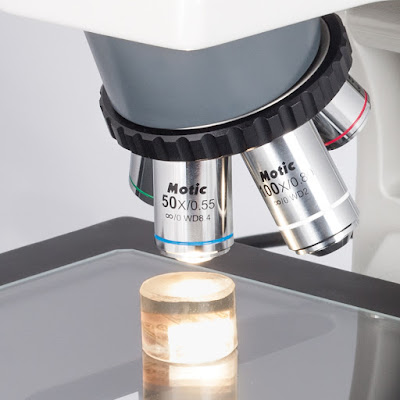
Opaque samples are treated with incident light. Take care of the correct selection of objectives without cover glass correction. The objective sleeve indicates this feature as a “0” (in the following case behind ∞, referring to the optical concept):
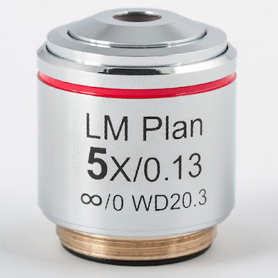
Motic Microscopes LM Plan 5x BF Objective from MMS Microscopes